Annalise Enterprise: decision-support AI for medical imaging
Team
Company | Institution
Category
Type
Project description
Medical imaging is the backbone of clinical medicine, informing diagnostic and patient management decisions across the healthcare system. The ever-growing number of medical examinations and worldwide decrease in staff challenges radiologists and healthcare providers to read more complex scans with decreasing resources while maintaining high standards of clinical excellence. Because of this, even well-resourced healthcare systems face significant imaging backlogs. Under-resourced healthcare systems with low clinician-to-patient ratios may miss opportunities for early disease detection due to long patient wait times.
AI can assist radiologists in critical triage and diagnostics, acting as “a second pair of eyes” in high-pressure, stressful conditions, helping to achieve the best patient outcomes. Annalise.ai’s advanced machine learning algorithms and software interface integrates seamlessly into their workflow, allowing them to treat critical patients sooner, reporting faster and more accurately.
Annalise Enterprise AI models were trained on almost 1 million unique, de-identified patient studies hand-labelled by >140 fully trained consultant radiologists, producing hundreds of millions of individual data points.
Annalise.ai AI tools serve to improve the accuracy of image interpretation, leading to better patient outcomes and enhanced workflow efficiencies in a world of ever-increasing demand for radiological services.
Proudly clinician-led, our patient-first approach comes from a deep understanding of the challenges faced in medical imaging. We fully understand the importance of our work and deeply care about the outcomes we work to support. OPT-PRM-100 Ver 0 Interaction Design Award 2023 Project Description
Description
Annalise.ai was founded in 2019 when we started researching, designing and building our first AI model for chest X-ray targeting a comprehensive list of clinical findings, compared to the industry standard of including only one or a few clinical findings. In 2021 already, we released our first regulatory-approved product, and were busy researching and designing the much more complex AI for non-contrast head CT.
Through iterative research and design, observing, interviewing and testing with international clinicians, we have understood their work’s incredible time pressure, unique operating environment, and fatiguing characteristics, including “eye miles” exerted scanning large visual fields and interacting with small, high-density interfaces.
Ergonomics, usability, efficiency and responsiveness are significant factors in any software tool’s adoption; a tool used constantly must be lightweight, low-friction, efficient and pleasing to sustain use and positively impact critical work, such as diagnostic imaging.
We want clinicians only to interact enough to inform diagnosis — the less interaction, the faster to critical information, the sooner to treat the next patient.
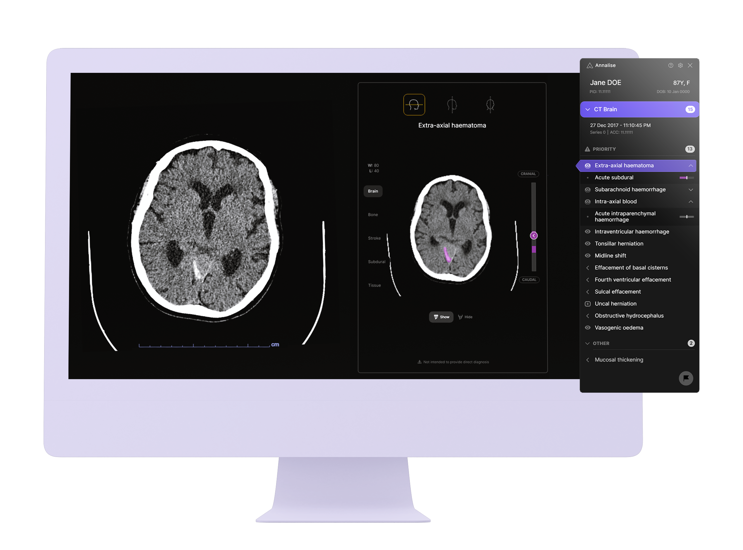
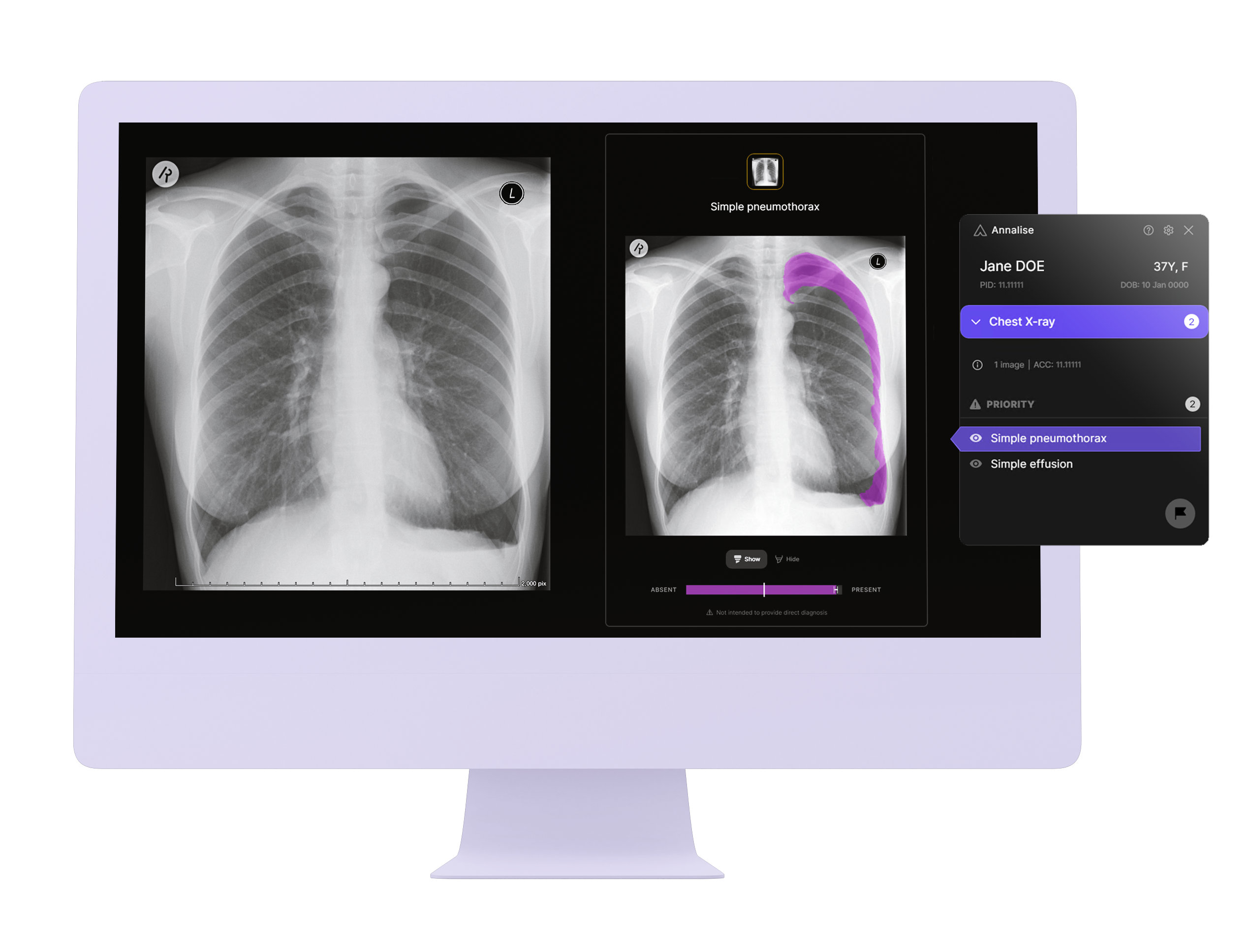
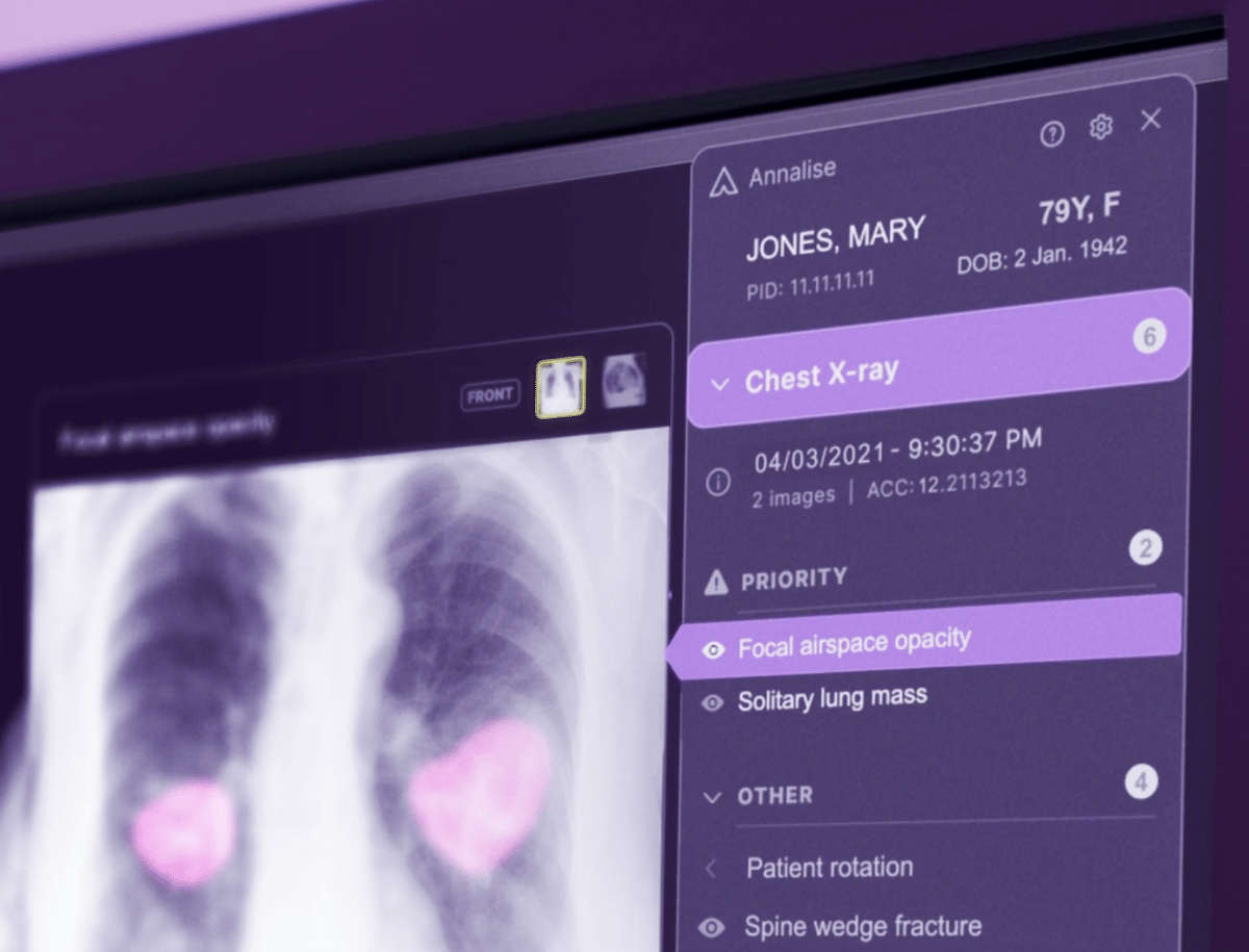
- The interface is carefully designed for progressive disclosure, expanding to reveal more detail as needed.
- Results are immediately available when clinicians open a study, expanded or collapsed to reduce cognitive biasing. Users can quickly scan the list and focus on the most concerning findings.
- Users quickly point at findings to expand the UI, highlighting the suspected finding on the patient image without further clicks or direct manipulation.
- To speed viewing in case of 3D scans, images are auto-straightened and use the best clinical view (axial, coronal, sagittal) on the critical slice showing the most significant extent of the abnormality.
- Each finding preview image is auto-adjusted according to the specific disease marker, something radiologists must constantly do as they search for abnormalities.
- In the case of 3D scans, our Slice Scrolling UI previews the spread of a finding. For more critical findings, clinicians can scroll to see precise contour changes showing how a finding impacts other anatomy.
- With feedback mode, users give feedback on AI findings and visualisations and can audit/tune AI sensitivity thresholds.
Annalise Enterprise modules detect up to 124 CXR and up to 130 CTB findings on the relevant input examinations. The AI algorithms were trained on over 782,000 unique CXR studies and over 229,000 non-contrast CTB studies, hand-labelled by >140 radiologists, generating >280 million CXR and >269 million CTB labels, respectively. Training datasets for Annalise Enterprise modules are amongst the world’s largest AI training datasets. OPT-PRM-100 Ver 0 Interaction Design Award 2023 Project Description
A major peer-reviewed retrospective multi-reader multi-centre validation study published in Lancet Digital Health (Jul-21) demonstrated that Annalise Enterprise CXR improved radiologist accuracy by 45% while speeding reporting time by 12%, averaged across all findings.
An observational, prospective study (BMJ, Dec-21) demonstrated improvement in radiologist reporting in hospital and community clinic settings: 1 in 30 cases had significant report changes, and 1 in 70 resulted in patient management changes.
Qualitative feedback from Annalise Enterprise users at a partnering site highlights that 90% of users think using the tool improves reporting accuracy for both CXR and CTB modules and is easy to use and learn in less than 10 cases for most users.
AI can assist radiologists in critical triage and diagnostics, acting as “a second pair of eyes” in high-pressure, stressful conditions, helping to achieve the best patient outcomes. Annalise.ai’s advanced machine learning algorithms and software interface integrates seamlessly into their workflow, allowing them to treat critical patients sooner, reporting faster and more accurately.
Annalise Enterprise AI models were trained on almost 1 million unique, de-identified patient studies hand-labelled by >140 fully trained consultant radiologists, producing hundreds of millions of individual data points.
Annalise.ai AI tools serve to improve the accuracy of image interpretation, leading to better patient outcomes and enhanced workflow efficiencies in a world of ever-increasing demand for radiological services.
Proudly clinician-led, our patient-first approach comes from a deep understanding of the challenges faced in medical imaging. We fully understand the importance of our work and deeply care about the outcomes we work to support. OPT-PRM-100 Ver 0 Interaction Design Award 2023 Project Description
Description
Annalise.ai was founded in 2019 when we started researching, designing and building our first AI model for chest X-ray targeting a comprehensive list of clinical findings, compared to the industry standard of including only one or a few clinical findings. In 2021 already, we released our first regulatory-approved product, and were busy researching and designing the much more complex AI for non-contrast head CT.
Through iterative research and design, observing, interviewing and testing with international clinicians, we have understood their work’s incredible time pressure, unique operating environment, and fatiguing characteristics, including “eye miles” exerted scanning large visual fields and interacting with small, high-density interfaces.
Ergonomics, usability, efficiency and responsiveness are significant factors in any software tool’s adoption; a tool used constantly must be lightweight, low-friction, efficient and pleasing to sustain use and positively impact critical work, such as diagnostic imaging.
We want clinicians only to interact enough to inform diagnosis — the less interaction, the faster to critical information, the sooner to treat the next patient.
- The interface is carefully designed for progressive disclosure, expanding to reveal more detail as needed.
- Results are immediately available when clinicians open a study, expanded or collapsed to reduce cognitive biasing. Users can quickly scan the list and focus on the most concerning findings.
- Users quickly point at findings to expand the UI, highlighting the suspected finding on the patient image without further clicks or direct manipulation.
- To speed viewing in case of 3D scans, images are auto-straightened and use the best clinical view (axial, coronal, sagittal) on the critical slice showing the most significant extent of the abnormality.
- Each finding preview image is auto-adjusted according to the specific disease marker, something radiologists must constantly do as they search for abnormalities.
- In the case of 3D scans, our Slice Scrolling UI previews the spread of a finding. For more critical findings, clinicians can scroll to see precise contour changes showing how a finding impacts other anatomy.
- With feedback mode, users give feedback on AI findings and visualisations and can audit/tune AI sensitivity thresholds.
Annalise Enterprise modules detect up to 124 CXR and up to 130 CTB findings on the relevant input examinations. The AI algorithms were trained on over 782,000 unique CXR studies and over 229,000 non-contrast CTB studies, hand-labelled by >140 radiologists, generating >280 million CXR and >269 million CTB labels, respectively. Training datasets for Annalise Enterprise modules are amongst the world’s largest AI training datasets. OPT-PRM-100 Ver 0 Interaction Design Award 2023 Project Description
A major peer-reviewed retrospective multi-reader multi-centre validation study published in Lancet Digital Health (Jul-21) demonstrated that Annalise Enterprise CXR improved radiologist accuracy by 45% while speeding reporting time by 12%, averaged across all findings.
An observational, prospective study (BMJ, Dec-21) demonstrated improvement in radiologist reporting in hospital and community clinic settings: 1 in 30 cases had significant report changes, and 1 in 70 resulted in patient management changes.
Qualitative feedback from Annalise Enterprise users at a partnering site highlights that 90% of users think using the tool improves reporting accuracy for both CXR and CTB modules and is easy to use and learn in less than 10 cases for most users.